Home
/ Foot And Leg Bones Diagram : Https Encrypted Tbn0 Gstatic Com Images Q Tbn And9gcrazglpnzcaleqxd2m9t0c Bntjai4l5gu5z3m G4ejh8gg5vwj Usqp Cau - Like already mentioned, the hindfoot is the posterior part of the foot.
Foot And Leg Bones Diagram : Https Encrypted Tbn0 Gstatic Com Images Q Tbn And9gcrazglpnzcaleqxd2m9t0c Bntjai4l5gu5z3m G4ejh8gg5vwj Usqp Cau - Like already mentioned, the hindfoot is the posterior part of the foot.
Foot And Leg Bones Diagram : Https Encrypted Tbn0 Gstatic Com Images Q Tbn And9gcrazglpnzcaleqxd2m9t0c Bntjai4l5gu5z3m G4ejh8gg5vwj Usqp Cau - Like already mentioned, the hindfoot is the posterior part of the foot.. The calcaneus, or heel bone: At the same time, the bones and joints of the leg and foot must be strong enough to support the body's weight while remaining. Its made up of 4 bones; The bones of the foot provide mechanical support for the soft tissues; Fpe medical review board a foot pain diagram is a great tool to help you work out what is causing your ankle and foot pain.
The anatomy of the leg and foot bones. The medial, larger bone of the lower leg. Rolling your ankle can cause a break in the knobby bumps at the end of the tibia and fibula. Chloe wilson bsc(hons) physiotherapy reviewed by: The leg is specifically the region between the knee joint and the ankle joint.
Leg Bone Wikipedia from upload.wikimedia.org This consists of five long metatarsal bones and five shorter bones that form the toes (phalanges). Let's review all of these bones one last time. The knee joint is the largest joint in the body and is primarily a hinge joint, although some sliding and rotation occur. In a typical foot the tibia is responsible for supporting about 85% of body weight. Bone diagram forehead (frontal bone) nose bones (nasals) cheek bone (zygoma) upper jaw (maxilla) lower jaw (mandible) breast bone (sternum) upper arm bone (humerus) lower arm bone (ulna) thigh bone (femur) collar bone (clavicle) toe bones (phalanges) ankle bones (tarsals) kneecap (patella) shin bone (tibia) calf bone (fibula) foot bones They can be divided into three groups: Quizzes on human skeletal system anatomy, bone anatomy, and bone markings. The talus, or ankle bone:
Moveable tissue) surround the true ankle and subtalar joints, binding the bones of the leg to each other and to those of the foot.
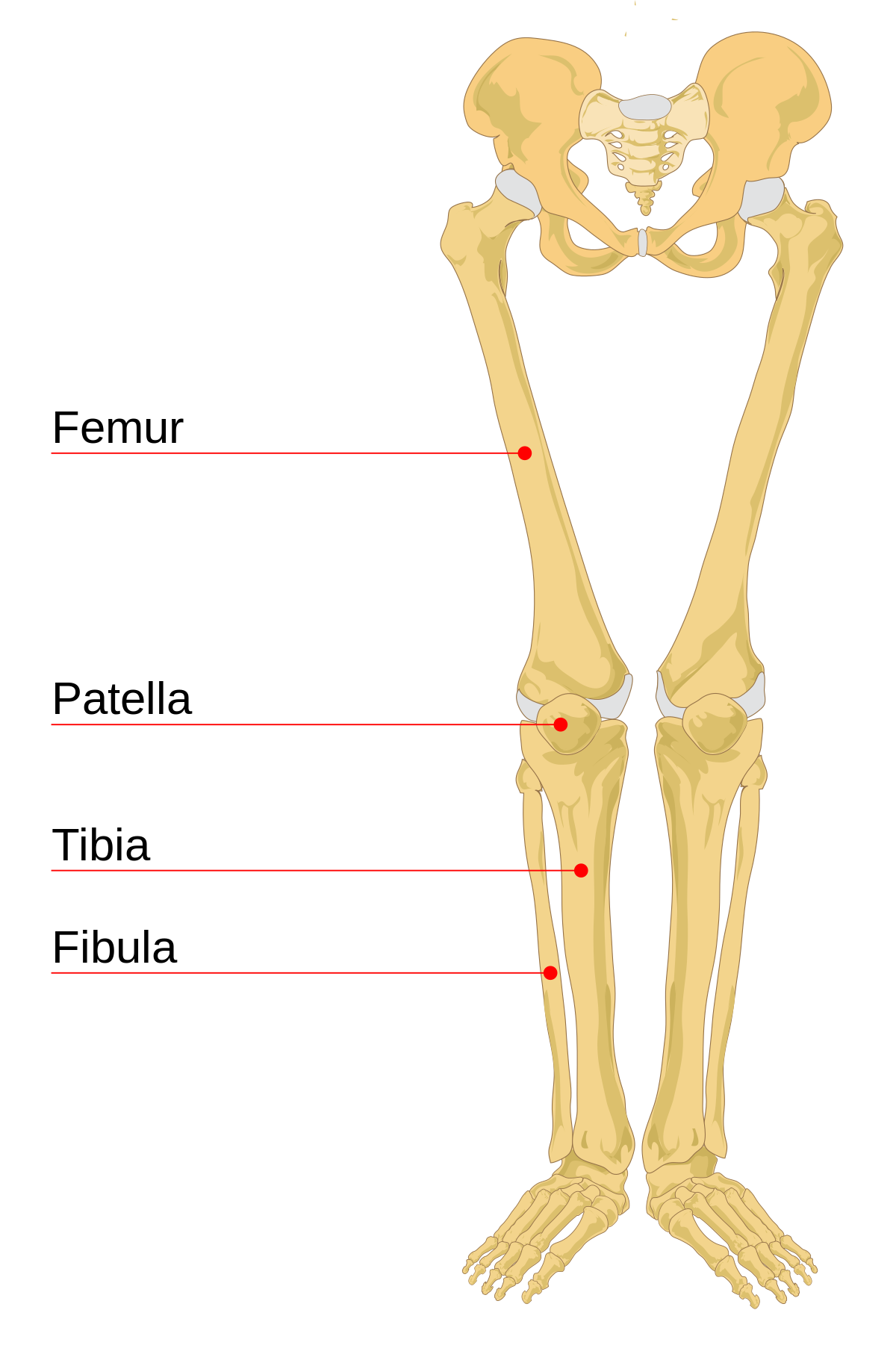
These are the main muscles that facilitate movement in the foot: Hol dir nike leg online. The bones of the leg are the femur, tibia, fibula and patella.the foot bones shown in this diagram are the talus, navicular, cuneiform, cuboid, metatarsals and calcaneus. Fpe medical review board a foot pain diagram is a great tool to help you work out what is causing your ankle and foot pain. The talus is the bone at the top of the foot. Helping the foot withstand the weight of the body whilst standing and in motion. Learn with flashcards, games, and more — for free. Like already mentioned, the hindfoot is the posterior part of the foot. When one looks at the anatomy of the foot, they would realize that the foot has a complex mechanical and structural architecture. Typically caused by trauma or injury to the foot or toe. Bones, muscles, tendons and nerves which will each give slightly different foot pain symptoms. Let's review all of these bones one last time. The bones of your leg and foot helped give you the ability to score that field goal.
Helping the foot withstand the weight of the body whilst standing and in motion. The talus, or ankle bone: Die heißesten sneaker releases online. The anatomy of the leg and foot bones. When one looks at the anatomy of the foot, they would realize that the foot has a complex mechanical and structural architecture.
Leg And Knee Anatomy Bones Muscles Soft Tissues Kenhub from thumbor.kenhub.com Tibia and fibula (long bones) the foot is connected to the body where the talus articulates with the tibia and fibula. At the same time, the bones and joints of the leg and foot must be strong enough to support the body's weight while remaining. The diagram of bones in the ankle and foot is given below: Tibialis posterior (supports the foot's arch) tibialis anterior (allows the foot to move upward) Die heißesten sneaker releases online. The seven tarsal bones are: These muscles work together to produce movements such as standing, walking, running, and jumping. They can be divided into three groups:
It connects with the tibia and fibula bones of the lower leg.
The medial, larger bone of the lower leg. Bones, muscles, tendons and nerves which will each give slightly different foot pain symptoms. This consists of five long metatarsal bones and five shorter bones that form the toes (phalanges). The diagram of bones in the ankle and foot is given below: Typically caused by trauma or injury to the foot or toe. The seven tarsal bones are: Distal to the ankle is the foot. The muscles that control the movements of the foot originate in the lower leg and are attached the bones in the foot with tendons. When one looks at the anatomy of the foot, they would realize that the foot has a complex mechanical and structural architecture. The tarsal bones work together as a group. This bone creates the lower portion of the ankle joint.; Related posts of diagram of leg bones bone anatomy elbow. At the same time, the bones and joints of the leg and foot must be strong enough to support the body's weight while remaining.
The calcaneus, talus, fibula, and tibia bones. The femur is the single bone of the thigh. The smaller lateral bone of the lower leg. Its main function is to allow for plantar flexion and dorsiflexion of the foot. Knee, leg, and foot (overview) how many times have a layman's language and anatomy ever matched?
1 from Bone diagram forehead (frontal bone) nose bones (nasals) cheek bone (zygoma) upper jaw (maxilla) lower jaw (mandible) breast bone (sternum) upper arm bone (humerus) lower arm bone (ulna) thigh bone (femur) collar bone (clavicle) toe bones (phalanges) ankle bones (tarsals) kneecap (patella) shin bone (tibia) calf bone (fibula) foot bones Below are slightly more detailed descriptions of the bones of the foot. At the same time, the bones and joints of the leg and foot must be strong enough to support the body's weight while remaining. The ankle joint is the shock absorber of the foot. Let's have a look at the bones of the ankle and foot. The bones of the foot provide mechanical support for the soft tissues; The smaller lateral bone of the lower leg. These are the main muscles that facilitate movement in the foot:
The way these bones fit together and interact with one another is very interesting.
Learn with flashcards, games, and more — for free. The anatomy of the leg and foot bones. It connects with the tibia and fibula bones of the lower leg. Ankle & lower leg anatomy. The tibia is one of the 2 bones that make up the leg. The proximal portion of the tibia is tibial plateau which acts as a cusp for the knee, the distal portion tapers into the medial malleoli and the concave surface which articulates with the talus at the ankle joint. The talus, or ankle bone: The tarsal bones work together as a group. The foot bones shown in this diagram are the talus, navicular, cuneiform, cuboid, metatarsals and calcaneus. The knee joint is the largest joint in the body and is primarily a hinge joint, although some sliding and rotation occur. Knee, leg, and foot (overview) how many times have a layman's language and anatomy ever matched? Tibia and fibula (long bones) though the tibia (commonly called the shin bone) is not a part of the foot, it plays an important role. #1 way to prevent lameness is to purchase a horse with good conformation.
Quizzes on human skeletal system anatomy, bone anatomy, and bone markings leg bones diagram. The bones of the foot provide mechanical support for the soft tissues;